Through this STSM, I aimed at gaining epertise in working with human intestinal organoids, to further enrich the Home Institution's expertise in developing more physiological relevant biomimetic in vitro intestinal models.

With regard to the accomplished practical work, initially, human-derived intestinal organoids were maintained in culture for 7 days (Figure 1) and subsequently passaged or differentiated into apical-out configuration (Figure 2), allowing me to become familiar with key aspects of their handling, differentiation, and maintenance.

Figure 1. Human jejunal organoids in culture for 7 days (Magnification: 4x).

Figure 2. Differentiated human jejunal organoids into apical-out configuration (Magnification: 40x). Another major goal of this STSM was to evaluate the intestinal permeability of exenatide-based delivery systems using this 3D intestinal model. Permeability studies were conducted by testing 1 µM of exenatide released from delivery systems, using human intestinal organoids differentiated into the apical-out configuration, after 24 hours.

In parallel, I acquired hands-on training in the development of monolayers derived from intestinal organoids—another key objective of this STSM. Organoids were enzymatically dissociated into single cells and seeded with different cells densities onto inserts (24-well plate format) coated with a Matrigel/Collagen I mixture (1:1). Media was changed every other day for 5 days and TEER values were measured to ensure the formation of a complete monolayer (Figure 3).

Figure 3. Monolayers-derived from human intestinal organoids seeded at different cell densities.

As an alternative approach to evaluate the efficacy of the delivery systems in the absence of human pancreatic islets, a bioluminescence resonance energy transfer (BRET)-based assay was employed to monitor the activation of the GLP-1 receptor (GLP-1R). This method enabled us to assess the functional engagement of the receptor by the released recombinant exenatide, providing valuable insights into the bioactivity of the delivery systems.

In summary, a new collaboration was established between the Home (i3S, Porto, Portugal) and Host Institution by my STSM focusing on the development of advanced in vitro intestinal models to be used as an alternative to animal models for drug testing. This transfer of knowledge contributes to the reduction of animal experiments used for drug testing and nanomedicines, in accordance with the goals of working group 1 of the CA IMPROVE. I have received specific training in culture of human intestinal organoids and in the establishment of monolayers-derived form intestinal organoids that will be applied at my Home Institution to improve the reproducibility and physiological relevance of the in vitro intestinal model.

Beyond scientific outcomes, the STSM contributed significantly to my personal and professional development. The opportunity to work in a new research environment abroad enriched my doctoral training, in line with the goals of working group 4 of the CA IMPROVE.

During my Short-Term Scientific Mission (STSM) at the Toxalim DR2 INRAE Institute in Toulouse, France, I worked on non-genotoxic carcinogens (NGTxCs)—chemicals that induce cancer without directly damaging DNA. In addition to advancing my theoretical understanding of these substances, I gained practical expertise in developing and analyzing advanced in vitro 3D co-culture models. Throughout this project, I worked under the mentorship of Dr. Marc Audebert. My primary focus during the STSM was to establish and characterize a 3D co-culture system using three different liver cell lines: HepaRG (human liver cells), h-TERT-HSC (hepatic stellate cells), and TPH-1 (monocytic cells).

The aim was to create a more complex, physiologically relevant model to study NGTxC-induced toxicity. Using magnetic beads (NanoShuttle-Grainer), I integrated these three cell types. The magnetic beads facilitate cell aggregation, promoting the formation of spheroids that more closely mimic the structural and functional complexity of human liver tissue. These beads are biocompatible, composed of gold, iron oxide, and Poly-L-Lysine, and they do not affect cell proliferation, viability, metabolism, oxidative stress, or the phenotype. Furthermore, they do not interfere with experimental techniques such as immunohistological labeling, fluorescence detection, HR microscopy, Western Blot, or PCR/qPCR.

Once the multi-cellular co-culture system was established, I characterized it in terms of cell viability, differentiation, and interactions, ensuring it was suitable for toxicological testing. The INRAE institute is a global leader in developing systems that replicate human physiology, particularly in the fields of toxicology and biomedical research. Their expertise in tissue engineering and in vitro technologies proved invaluable throughout this process.

After learning how to maintain the co-culture system, I focused on applying confocal microscopy to analyze fixed 2D and 3D cell samples. I first learned how to fix and stain both monolayer cells and multicellular spheroids specifically for confocal imaging. Using advanced staining techniques, I labeled the spheroids with specific markers and fluorochromes to visualize cellular morphology, protein localization, and interactions within the co-culture system. The host institution’s extensive experience with confocal microscopy enabled high-resolution imaging of cellular interactions and structural changes, allowing for detailed analysis of these complex biological systems.

Once the co-culture models were established and characterized, I exposed the spheroids to selected NGTxCs and non-carcinogenic chemicals (as controls) to evaluate their effects. The exposure lasted for 24 and 96 hours (with repeat treatments) using five different compounds: etoposide (genotoxic), colchicine (genotoxic), KBrO3, D-mannitol, and a control. For each treatment, four spheroids were exposed to five concentrations of each compound in 10-fold dilutions. All experiments were performed in triplicate.

To assess the impact of the treatments, I first observed structural changes in the treated spheroids using confocal microscopy. This high-resolution imaging provided insights into cellular morphology, protein localization, and the interactions between cells within the co-culture system. I used a panel of five cell markers—DAPI (for nuclei), pH3 (for mitotic cells), GammaH2A.X (for double strand breaks), HMOX1 (for oxidative stress response), and IL-6 (for inflammatory response)—to characterize cellular responses to the treatments.

Additionally, we collected samples for further transcriptomic analysis, which will take place at the National Institute of Biology (NIB). This analysis will assess changes in gene expression triggered by NGTxCs, identifying the molecular pathways involved in NGTxC-induced carcinogenesis. These insights are critical for understanding the cellular mechanisms driving NGTxC toxicity.

The outcomes of the STSM directly support the objectives of the COST Action IMPROVE by:

In conclusion, the STSM successfully met its scientific, technical, and collaborative goals. It contributed meaningfully to the development of New Approach Methodologies (NAMs) and established a strong basis for long-term international cooperation under the framework of the COST Action IMPROVE.

Through this STSM, the applicant aimed at learning the methods available at the Host Institution for the bioprinting of alginate-based bioinks embedding hepatic cells, as well as techniques for evaluating the bioprinted constructs and the cells embedded in them. Briefly, during the STSM, the applicant had opportunity to perform the bioprinting of alginate and gelatin bioinks and test the effect of different crosslinking conditions on the stability of the bioprinted constructs. The referred bioinks were also used for embedding hepatic cells, mainly hepatocytes (HepG2 cell line) and hepatic stellate cells (LX-2 cell line). Cells embedded in the scaffolds were analysed using different techniques. Moreover, the applicant was also able to test the 3D bioprinting of a different bioink composed by collagen type I and alginate. The system used for bioprinting at the Host Institution is presented in Figure 1, and representative pictures of the printed scaffolds are shown in Figure 2.

Overall, the initial work plan proposed in the STSM was achieved, as the applicant was able to gain extensive hands-on experience on 3D bioprinting of alginate-based bioinks for creating in vitro liver models that will be essential for her work at the Home Institution iS3 in Porto, Portugal.

Within the STSM which took place at the IRCCS Fondazione Policlinico Universitario A. Gemelli from the 5th of April until the 5th of June 2024, I took a 2-month hands-on-learning visit at the Organoids Research Core Facility under the supervision of Professor Claudio Sette.

I learned a lot about the culture and handling of high-grade serous ovarian cancer organoids and ovarian cancer cell lines, as well as different advanced experimental techniques such as western-blot, proliferation and viability assays.

I have had the opportunity to advance the research associated with my PhD project, being able to acquire valuable data and results that will undoubtably enhance the potential of the compound we are developing.

Moreover, this STSM also contributed to the advance my academic and professional growth.

I can say with certainty that I have broadened my knowledge about in vitro 3D models, particularly organoids.

The visit to IRCCS Fondazione Policlinico Universitario A. Gemelli was an extremely pleasant experience and I would not hesitate to do it again.

Figure: Three different high-grade serous ovarian cancer organoids cultured and handled during my experience.

I am Paolo Signorello, a PhD student at the Department of Information Engineering, University of Pisa, and a member of Centro 3R, Italy. Thanks to the Short-Term Scientific Mission (STSM), I spent four months (from 01/02/2024 to 31/05/2024) at “i3S - Instituto de Investigação e Inovação em Saúde da Universidade do Porto”, under the supervision of Professor Bruno Sarmento. During this period, I developed a magnetic nanoparticle-based formulation and tested it in an advanced in vitro intestinal model1 for potential use in drug delivery or hyperthermic therapy targeting the intestine.

Figure 1: Badge at i3S.

This model consisted, from top to bottom, of a multilayer co-culture using transwells for 24-well plates, which included four different cell types: two types for the epithelial layer, fibroblasts encapsulated in a hydrogel to ensure the development of the extracellular matrix in the apical compartment, and endothelial cells in the basolateral compartment. This configuration was designed to mimic the complex structure of the intestinal barrier.

First, I functionalized magnetic nanoparticles with a mucoadhesive biological material and tested them for stability over time and at different pH levels using a Dynamic Light Scattering (DLS) instrument.

Subsequently, I conducted a viability assay to evaluate the toxicity of the magnetic nanoparticles on all cell types. After 21 days of developing the model, I tested for any biological differences that may occur in terms of permeability and nutrient absorption, both in the presence and absence of the magnetic nanoparticle-based formulation.

I am very grateful to the COST Action IMPROVE for supporting this period abroad at i3S, as it significantly enhanced my knowledge of nanomaterials, cytotoxicity, the development of complex in vitro models, and co-culture techniques.

The atmosphere at i3S is very welcoming and inclusive. Furthermore, Professor Sarmento’s team is very proactive in building new working groups and fostering networking opportunities. This experience will allow me to engineer better, more advanced models with improved predictivity, in line with the 3Rs Principles.

Figure 2: A) Nanoparticles formulation and B) Dynamic Light Scattering (DLS) instrument.

[1] Ferreira B, Barros AS, Leite-Pereira C, Viegas J, das Neves J, Nunes R, Sarmento B. Trends in 3D models of inflammatory bowel disease. Biochim Biophys Acta Mol Basis Dis. 2024 Mar;1870(3):167042. doi: 10.1016/j.bbadis.2024.167042. Epub 2024 Jan 29. PMID: 38296115.

Within the STSM which took place at Swansea University Medical School from the 15th of April until the 19th of April 2024, I took an intensive 4–5-day hands-on-learning visit at the Department of the In Vitro Toxicology Group under the supervision of Professor Shareen Doak.

I learned a lot about the handling of nanomaterials in the field of in vitro toxicology, different kinds of exposure techniques of advanced (3D) in vitro cell models, techniques for studying nanomaterial cell internalization, and was introduced to the approach they use for assessing the nanoparticle toxicity.

I have successfully learned about the safe handling of nanomaterials (especially when it comes to dealing with waste that contains nanomaterials), I have seen different kinds of exposure techniques for both 2D and advanced 3D cell models and gotten to know the method used for nanomaterial cell internalization (TEM).

I have had a chance to see and try two additional methods for preparing spheroids not used at our institute – the Institute of Biology, Ljubljana, Slovenia.

What is even more important I have learned the comprehensive approach of nanoparticle toxicity assessment from preparing the suspension of nanoparticles, assessing their characteristic, and performing sonication, to exposing 2D and 3D cell models to nanoparticles using different exposure systems.

I can say with certainty that I have broadened my knowledge about nanomaterial handling, nanosafety, and different approaches in toxicology studies for assessing nanomaterial-based toxicity on 2D and advanced in vitro models. The visit to Swansea University was an extremely pleasant experience and I would not hesitate to do it again.

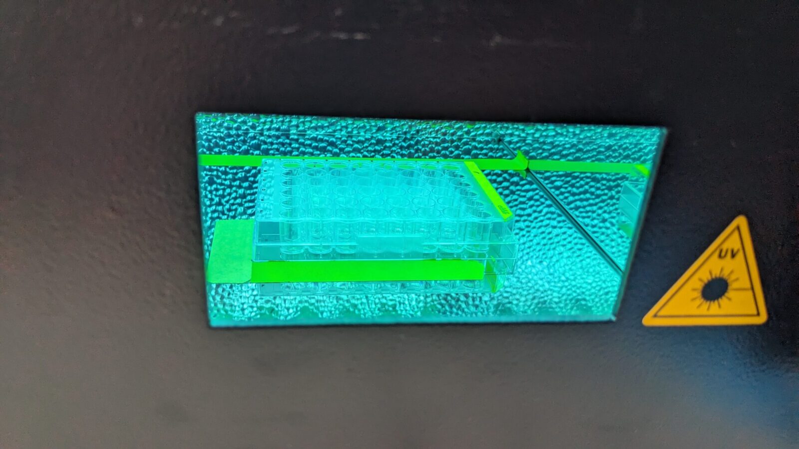
Dr. Eva Jablonská (University of Chemistry and Technology in Prague, Czech Republic) had a great opportunity to spend more than two months (15.1.-22.3.2024) at the Dermatotoxicology Study Centre, German Federal Institute for Risk Assessment (BfR) in Berlin. During the STSM granted by CA IMPROVE, she tested natural extracts from marine algae for photoprotective properties. Specifically, she irradiated human dermal fibroblasts with UV light and tested whether these extracts could prevent cellular damage.

Overall, the atmosphere at BfR was very welcoming and supportive. Additionally, the discussion about methods of genotoxicity testing were very fruitful.

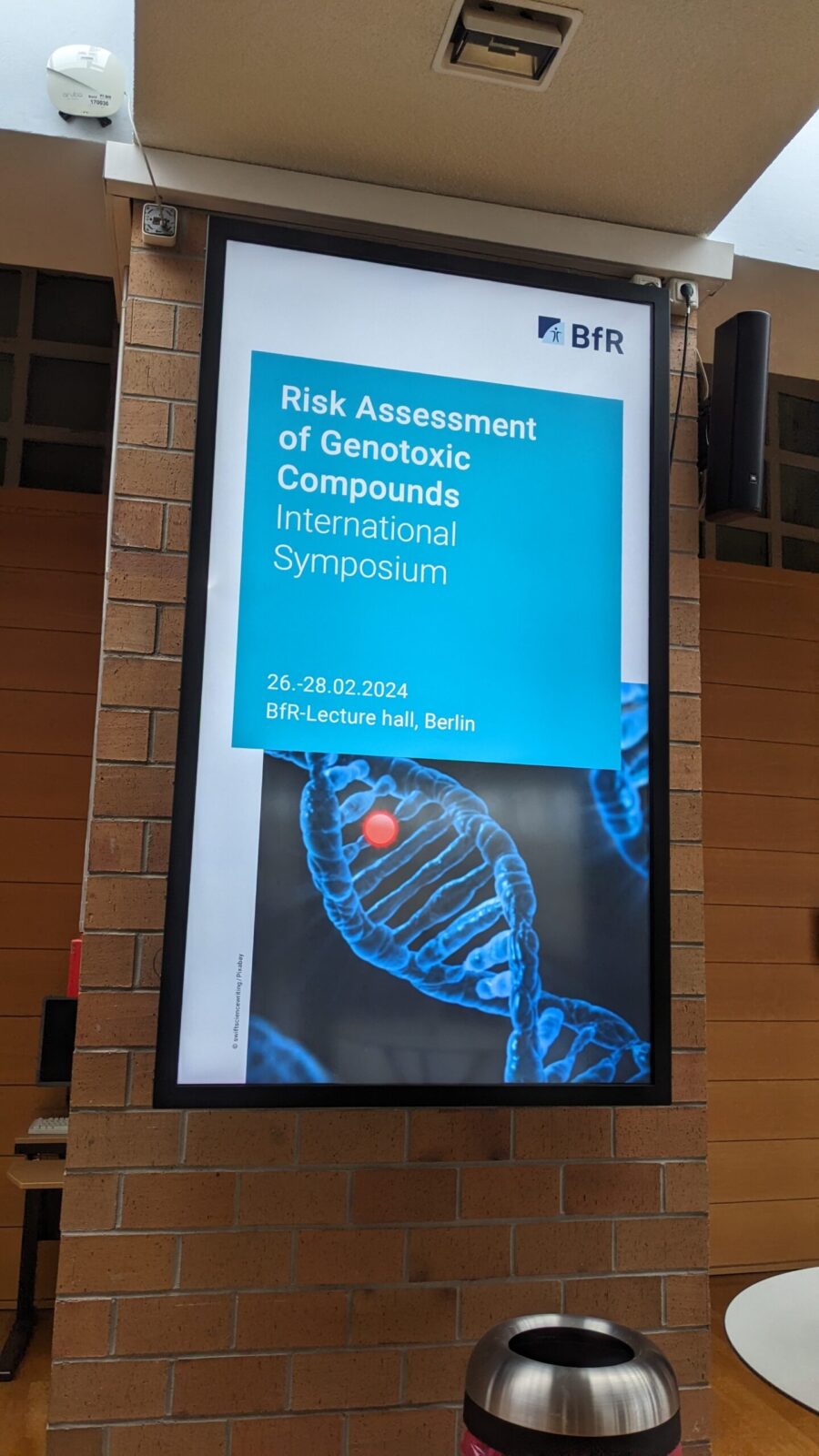
Moreover, the grantee was fortunate to attend a three-day symposium on genotoxicity held by BfR, where quantitative approaches towards genotoxicity assessment were discussed.

usercross